Exercise 15 histology of nervous tissue – Exercise 15: Histology of Nervous Tissue embarks on an illuminating exploration of the intricate structure and function of the nervous system. This captivating narrative unravels the mysteries of the brain and nerves, revealing the cellular components, structural organization, and functional aspects that orchestrate our thoughts, actions, and sensations.
As we delve into the realm of nervous tissue histology, we uncover the specialized cells, intricate networks, and remarkable adaptations that underpin the nervous system’s extraordinary capabilities. Prepare to be amazed as we unravel the secrets of this vital organ system, shedding light on its role in our physical and cognitive experiences.
Histological Techniques

The preparation of nervous tissue for microscopic examination involves specialized histological techniques. These techniques include:
- Fixation:Preserves tissue structure by cross-linking proteins.
- Embedding:Encases tissue in a support medium for sectioning.
- Sectioning:Creates thin slices of tissue for examination.
- Staining:Enhances tissue components for visualization.
Specific stains used in nervous tissue histology include:
- Hematoxylin and eosin (H&E):General stain for visualizing cell nuclei and cytoplasm.
- Luxol fast blue:Stains myelin sheaths blue.
- Bodian’s silver stain:Impregnates neurons with silver, highlighting their morphology.
Cellular Components of Nervous Tissue

Nervous tissue consists of two main cell types:
- Neurons:Excitable cells that transmit electrical and chemical signals.
- Glial cells:Support and protect neurons.
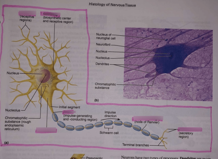
Neurons have a cell body (soma), dendrites (input processes), and an axon (output process). Glial cells include astrocytes, oligodendrocytes, microglia, and ependymal cells, each with specific functions in supporting neuronal activity.
Structural Organization of Nervous Tissue
Nervous tissue is organized hierarchically:
- Cells:The basic units of nervous tissue.
- Tissues:Groups of cells with similar functions, such as gray matter and white matter.
- Organs:Structures composed of multiple tissues, such as the brain and spinal cord.
- Nervous system:The entire network of nervous tissue.
The nervous system is divided into the central nervous system (CNS), consisting of the brain and spinal cord, and the peripheral nervous system (PNS), consisting of nerves and ganglia.
Functional Aspects of Nervous Tissue
Nervous tissue exhibits electrical and chemical properties that enable nerve impulse transmission:
- Electrical properties:Neurons have a resting membrane potential and generate action potentials, which are electrical signals that travel along axons.
- Chemical properties:Neurotransmitters are chemicals released at synapses that transmit signals between neurons.
The combination of electrical and chemical properties allows for rapid and efficient communication within the nervous system.
Comparative Histology of Nervous Tissue
Nervous tissue histology varies across species:
- Brain size:Humans have a larger brain size compared to other mammals, reflecting increased cognitive abilities.
- Neuronal density:The number of neurons per unit volume varies, affecting processing capabilities.
- Glial cell distribution:The ratio of glial cells to neurons influences nervous system efficiency.
These differences reflect evolutionary adaptations and functional specializations in different species.
Clinical Applications of Nervous Tissue Histology
Histological techniques are used in the diagnosis and treatment of neurological disorders:
- Brain biopsies:Examination of tissue samples for diagnosis of conditions such as tumors and infections.
- Neuropathology:Study of nervous tissue abnormalities associated with diseases like Alzheimer’s and Parkinson’s.
- Tissue engineering:Development of artificial neural tissue for transplantation and repair.
Histological analysis provides valuable insights into the pathology and treatment of neurological disorders.
Future Directions in Nervous Tissue Histology: Exercise 15 Histology Of Nervous Tissue

Emerging technologies and research areas in nervous tissue histology include:
- Advanced imaging techniques:High-resolution microscopy and computational analysis for detailed tissue visualization.
- Molecular profiling:Identification of genetic and molecular markers associated with neurological disorders.
- Stem cell-based therapies:Development of new treatments for neurological conditions using stem cells.
These advancements promise to enhance our understanding and treatment of nervous system disorders.
General Inquiries
What are the key histological techniques used to prepare nervous tissue for microscopic examination?
Common histological techniques include paraffin embedding, frozen sectioning, and Golgi staining. Paraffin embedding provides thin sections for general histological examination, while frozen sectioning allows for rapid processing and preservation of delicate structures. Golgi staining selectively impregnates neurons, revealing their intricate morphology.
What are the major cellular components of nervous tissue?
Nervous tissue consists of neurons, glial cells (astrocytes, oligodendrocytes, microglia), and ependymal cells. Neurons are the primary functional units, transmitting electrical and chemical signals. Glial cells provide support, insulation, and protection for neurons.
How is nervous tissue organized structurally?
Nervous tissue exhibits a hierarchical organization, from cells to organs. Neurons form synapses to create neural circuits within the central nervous system (brain and spinal cord) and peripheral nervous system (nerves and ganglia). These circuits are organized into functional regions, such as the cerebral cortex and cerebellum.