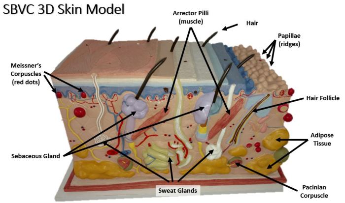
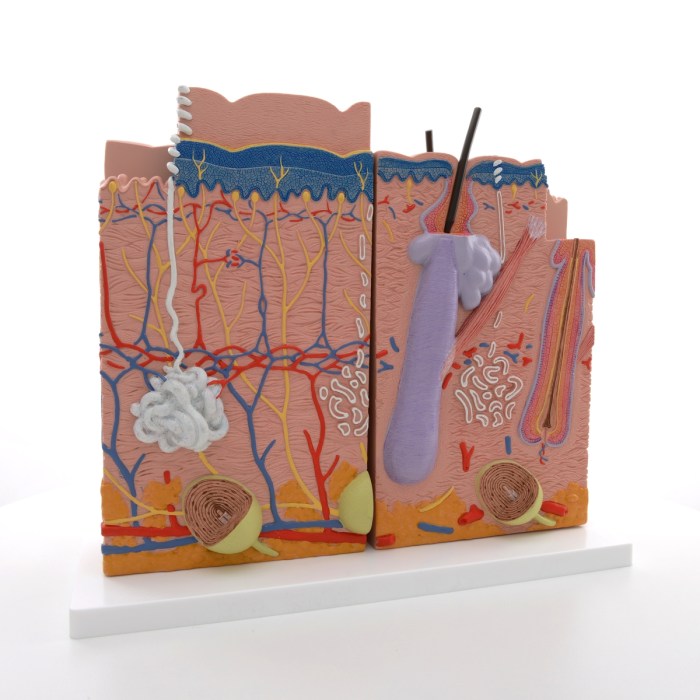
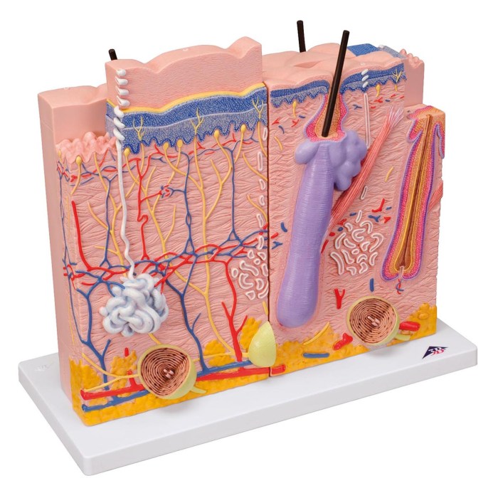
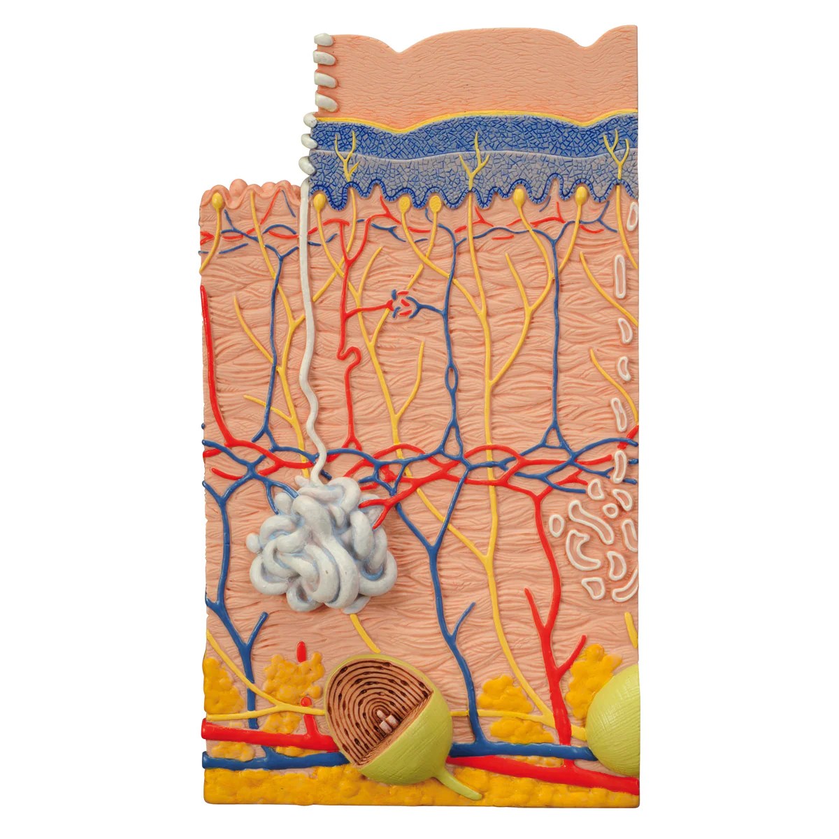
J16 3 part skin model labeled – Unveiling the J16 3-Part Skin Model Labeled, an intricate representation of the human skin’s structure, we embark on a captivating journey into the realm of dermatology.
This meticulously crafted model grants us an unparalleled opportunity to delve into the intricacies of the skin’s layers, their functions, and their significance in maintaining our overall health and well-being.
Anatomical Layers of J16 3-Part Skin Model
The J16 3-part skin model provides a detailed representation of the three distinct layers of human skin: the epidermis, dermis, and hypodermis. Each layer serves specific functions and exhibits unique characteristics.
Epidermis
- The epidermis is the outermost layer of the skin, responsible for providing a protective barrier against external elements.
- It is composed of multiple layers of keratinized cells, with the outermost layer being composed of dead cells.
- The epidermis is responsible for producing melanin, which provides skin pigmentation and protection from ultraviolet radiation.
Dermis
- The dermis is the middle layer of the skin, providing strength and elasticity.
- It contains a network of collagen and elastin fibers, along with blood vessels, nerves, and hair follicles.
- The dermis provides nourishment and support to the epidermis and aids in temperature regulation.
Hypodermis
- The hypodermis is the innermost layer of the skin, composed primarily of adipose tissue.
- It acts as an insulator, providing thermal insulation and cushioning for the body.
- The hypodermis also serves as a storage site for energy reserves.
| Layer | Thickness | Cell Types | Functions |
|---|---|---|---|
| Epidermis | 0.04-0.12 mm | Keratinocytes, melanocytes, Langerhans cells | Protection, pigmentation, barrier against pathogens |
| Dermis | 1-4 mm | Fibroblasts, macrophages, mast cells | Strength, elasticity, nourishment, temperature regulation |
| Hypodermis | Variable | Adipocytes | Insulation, cushioning, energy storage |
Epidermal Structures and Functions

The epidermis, the outermost layer of the skin, plays a crucial role in protecting the body from external factors. It comprises five distinct layers, each with specific functions:
Epidermal Layers
- Stratum Corneum:The outermost layer, composed of dead, keratinized cells, provides a waterproof barrier against external threats.
- Stratum Lucidum:Present only in thick skin, this thin, translucent layer helps enhance the skin’s barrier function.
- Stratum Granulosum:Consists of cells that produce and store granules containing antimicrobial peptides, contributing to skin defense.
- Stratum Spinosum:The thickest layer, composed of spiny-shaped cells that provide strength and resilience to the skin.
- Stratum Basale:The deepest layer, attached to the underlying dermis, contains stem cells that continuously divide to replace lost epidermal cells.
Keratinization
Keratinization is a vital process in which epidermal cells gradually transform into keratin-filled, dead cells. As they move towards the skin’s surface, they undergo biochemical changes that result in the formation of a protective layer, the stratum corneum. Keratinization plays a crucial role in maintaining skin integrity, preventing dehydration, and protecting against external insults.
Dermal Structures and Functions

Beneath the epidermis lies the dermis, a thicker and more complex layer that provides structural support and nourishment to the skin. Composed primarily of connective tissue, the dermis is a dynamic and metabolically active region.
The dermis is composed of two distinct layers:
- Papillary layer:The outermost layer, directly beneath the epidermis, is thin and highly vascularized. It contains numerous small blood vessels, nerve endings, and dermal papillae, which extend into the epidermis and provide nourishment and support to the overlying cells.
- Reticular layer:The thicker and deeper layer, composed of dense connective tissue, collagen and elastin fibers. These fibers provide strength and flexibility to the skin, allowing it to withstand stretching and recoil.
Blood Vessels
The dermis is richly supplied with blood vessels, including arteries, capillaries, and veins. These vessels provide nourishment to the skin cells and help regulate body temperature.
Hair Follicles
Hair follicles are embedded within the dermis. Each follicle consists of a hair shaft, hair root, and sebaceous gland. Sebaceous glands secrete an oily substance that lubricates the hair and skin.
Sweat Glands
Sweat glands are coiled tubular structures found in the dermis. They produce sweat, which helps regulate body temperature and eliminate waste products.
Nerve Endings, J16 3 part skin model labeled
The dermis contains numerous nerve endings that transmit sensory information to the brain. These nerve endings include touch receptors, temperature receptors, and pain receptors.
Hypodermal Structures and Functions

The hypodermis, also known as the subcutaneous layer, is the deepest layer of the skin. It lies beneath the dermis and is composed of loose connective tissue, fat cells (adipocytes), and blood vessels.
The J16 3 Part Skin Model Labeled provides an in-depth look at the anatomy of the skin. If you’re interested in brushing up on your US history, check out the US History STAAR Study Guide . It’s a great resource for students preparing for the STAAR exam.
Coming back to the J16 3 Part Skin Model Labeled, it’s an essential tool for students and medical professionals alike.
The hypodermis plays several important roles, including:
Insulation
- The fat cells in the hypodermis help to insulate the body, preventing heat loss.
- This is especially important in cold climates, where a thick layer of subcutaneous fat can help to keep the body warm.
Energy Storage
- The fat cells in the hypodermis also store energy in the form of triglycerides.
- This energy can be released when needed, such as during periods of fasting or exercise.
Cushioning
- The loose connective tissue in the hypodermis helps to cushion the body and protect it from injury.
- This is especially important in areas that are prone to impact, such as the buttocks and thighs.
Clinical Significance
The hypodermis can be affected by a number of conditions, including:
- Cellulitisis an infection of the hypodermis that can cause redness, swelling, and pain.
- Lipodystrophyis a condition that causes the loss of fat from the hypodermis. This can lead to a number of problems, including insulin resistance and diabetes.
Clinical Applications of the J16 3-Part Skin Model

The J16 3-part skin model is a valuable tool in dermatological research and education. It provides a realistic representation of the skin’s anatomy, allowing researchers and students to study skin structure and function in detail.The model has been used to investigate a wide range of skin diseases, including acne, psoriasis, and eczema.
It has also been used to study the effects of cosmetic procedures, such as laser therapy and chemical peels. Additionally, the model has been used to develop new wound healing therapies.
Studying Skin Diseases
The J16 3-part skin model has been used to study the pathogenesis of various skin diseases. By recreating the skin’s structure and function, researchers can observe how different factors contribute to the development of these diseases. For example, the model has been used to study the role of inflammation in acne and the role of immune dysfunction in psoriasis.
Cosmetic Procedures
The J16 3-part skin model has also been used to study the effects of cosmetic procedures. By applying different treatments to the model, researchers can observe how they affect the skin’s structure and function. For example, the model has been used to study the effects of laser therapy on wrinkles and the effects of chemical peels on acne scars.
Wound Healing
The J16 3-part skin model has been used to develop new wound healing therapies. By recreating the skin’s wound healing process, researchers can test the effectiveness of different treatments. For example, the model has been used to study the effects of growth factors on wound healing and the effects of antimicrobial agents on wound infection.
User Queries: J16 3 Part Skin Model Labeled
What are the three layers of the J16 3-Part Skin Model Labeled?
The three layers are the epidermis, dermis, and hypodermis.
What is the function of the epidermis?
The epidermis protects the body from external factors and prevents water loss.
What structures are found in the dermis?
The dermis contains blood vessels, hair follicles, sweat glands, and nerve endings.